Secondary Lesions Examples
Introduction to dermatology Skin lesions secondary excoriation 1. primary and secondary lesions
Image Gallery: Secondary Skin Lesions | Clinician's Brief
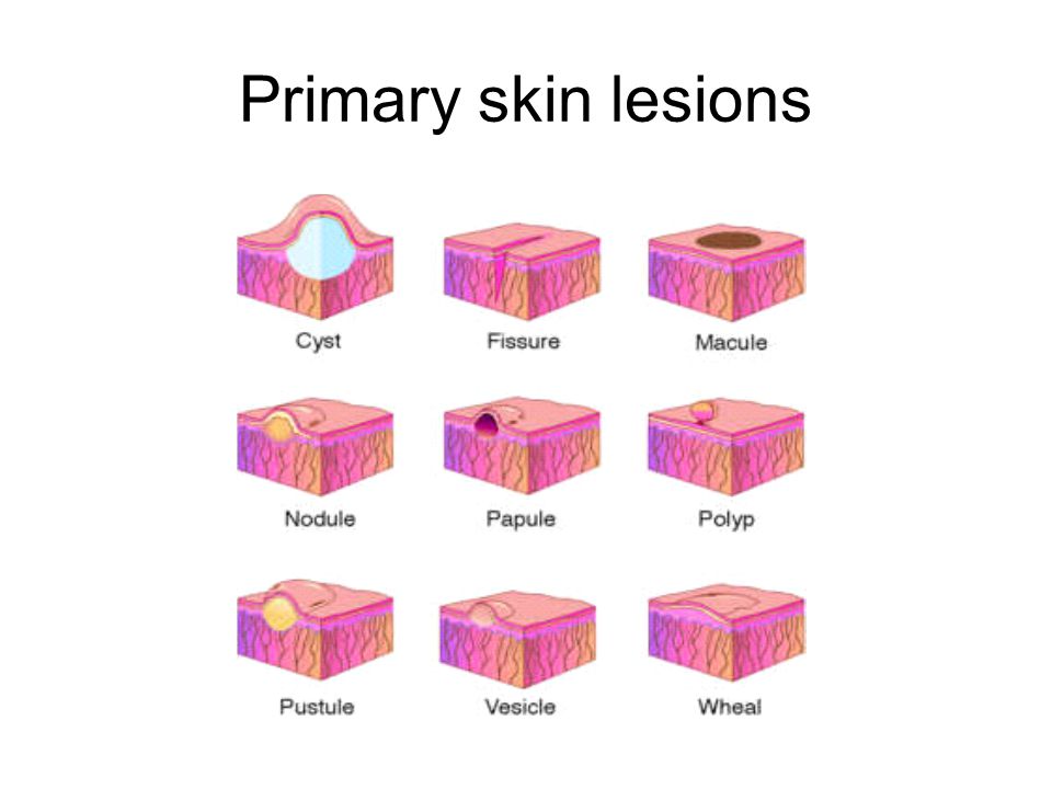
Examples of primary and secondary skin lesions. Lesions skin primary description lesion macule patch raised rashes pediatrics common medical study ppt powerpoint presentation Integumentary system alterations lesions skin lesion common figure basicmedicalkey characteristics
Primary lesions
Lesion dermatology bulla circumscribed filled elevated lesions primary fluid size greater thanAlterations in the integumentary system Secondary skin lesionsImage gallery: secondary skin lesions.
Lesions common malignant growths miiskinLesions secondary Lesions morphology dermatology conditions describing eczemaLesions lesion anatomy microbiota infections acne macule papule crust cyst vesicle wheal ulcer layers openstax secondary integumentary bacteria.

Anatomy and normal microbiota of the skin and eyes · microbiology
Image gallery: secondary skin lesionsSkin lesions secondary collarette Skin lesions nursing macule oral papule vs nodule dental medical dermatology school cyst primary papules lesion pustule secondary vesicle pustulesStudy medical photos: description of primary skin lesions.
Skin lesions: types, pictures & preventionPrimary and secondary lesions of the oral cavity Lesions cutaneous urticaria.


Primary and Secondary Lesions of the Oral cavity

Primary lesions

Image Gallery: Secondary Skin Lesions | Clinician's Brief

1. Primary and Secondary Lesions | Cutaneous Conditions | Skin

Skin Lesions: Types, Pictures & Prevention

Image Gallery: Secondary Skin Lesions | Clinician's Brief

Examples of primary and secondary skin lesions. | Download Table

Study Medical Photos: Description Of Primary Skin Lesions

Anatomy and Normal Microbiota of the Skin and Eyes · Microbiology

Introduction to Dermatology | The Basics | Describing Skin Lesions
